at Toray Research Center, Inc. and National Institute of Materials and Chemical Research
<1983 - 1997>
Analytical Software for Transmission-Mode Small Angle X-ray ScatteringPublication List
We developed a transmission-mode SAXS system and applied it to SAXS measurements on a variety of industrial materials, e.g., aluminum fiber, carbon fibers, and hard-elastic polypropylene films. With the SAXS system, we determined size distributions of aluminum particles, void size distributions in carbon fibers, and long periods and lamellar thicknesses of hard-elastic polypropylene films to discuss the structural parameters in terms of manufacturing conditions. In particular, lamellar structures of the hard-elastic polypropylene films, revealed by the SAXS analysis, were verified by transmission electron microscopy observations.
SAXS functions of various distorted multilayers (MLs) in, e.g., (1) lamellar structures of polymers, (2) microphase-separated structures of block copolymers and polymer blends, and (3) semiconductor and metal superlattices were formulated. The following structural modifications were considered: finiteness of the ML structure, paracrystalline distortions, layer-thickness distribution, layer curvature, transitional zone at the interface, and electron-density fluctuation. For each defect, characteristic features of the SAXS pattern were elucidated. A methodology for precise characterization of MLs by SAXS was proposed.
Reflection-Mode Small Angle X-ray Scattering and Its ApplicationsPublication List
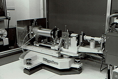
For structural determination of long-periodic layered materials such as Langmuir-Blodgett (LB) films and semiconductor superlattices (SLs), we designed a high-resolution reflection-mode small angle X-ray scattering (SAXS) setup based on the Kratky camera. The optical setup was assembled by Mr. Osamu Hirashima of Rigaku Corporation (shown in the upper figure), installed on a rotating-anode X-ray generator, and connected to a superminicomputer via a control unit. We also developed software for the device control and data analysis. The measurements for LB films and semiconductor SLs with our SAXS system demonstrated its superiority in performance over conventional SAXS apparatuses.
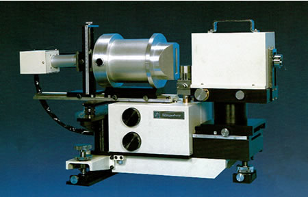
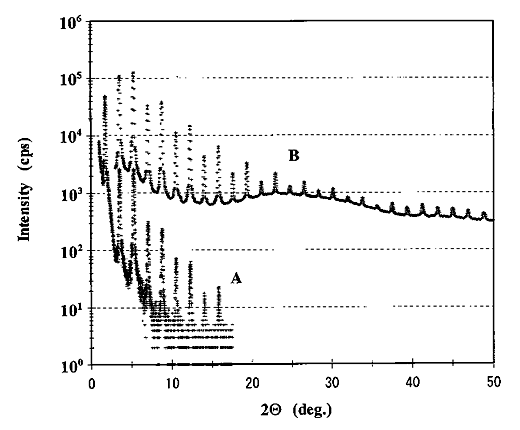
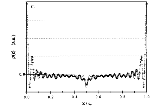
The SAXS intensities observed from LB films of lead stearate prepared under different conditions were analyzed in terms of lattice distortions by using the microstrain and paracrystalline models to correlate the distortions with the preparation conditions. For an LB film of cadmium arachidate, structural changes on heating and cooling were investigated, and melting behaviors of the organic thin film were clearly revealed. From an LB multilayer (21 layers) of cadmium stearate, high-quality SAXS data were obtained with the reflection-mode SAXS system (denoted as A in the middle figure) and a Bragg-Brentano setup (as B), and both scattering data were scaled and combined. As shown in the lower figure, the electron density (ED) profile in the stacking direction was derived from the Fourier synthesis of the combined intensities with the aid of semi-empirical molecular orbital calculations. In the ED profile, the outermost peaks, flat parts, and central valley represent the cadmium atoms, hydrocarbon chains, and a vacancy around the methyl terminals, respectively. I (Y.S.) wrote a thesis on these studies, submitted it to Department of Polymer Chemistry, Tokyo Institute of Technology, and successfully received a doctorate. The reflection-mode SAXS setup is now commercially available from Rigaku Corporation and has been used in academes and industries around the world.
A Point-Focusing Small Angle X-Ray Scattering Camera Using a Doubly Curved Monochromator of a W/Si Multilayer and Image Reconstruction by the Maximum Entropy MethodPublication List
We developed a point-focusing SAXS camera using a doubly curved monochromator of a W/Si multilayer in collaboration with Mr. Yuji Kobayashi and Mr. Katsunari Sasaki of Rigaku Corporation. The setup gives a very intense point beam. The performance of the SAXS camera was demonstrated by time-resolved measurements for liquid crystals (as a collaboration with Dr. Robert V. Law, present affiliation: Imperial College, UK). A combined use of the SAXS setup and two-dimensional data treatment based on the maximum entropy method has enabled us to observe SAXS images from polyethylene films by only 3 second exposure. The SAXS camera, adapted to be more compact, is commercially available as the NANO-Viewer from Rigaku Corporation.